2020-10-20

complement system -Is complement system innate or adaptive immunity?


Is the complement system innate or adaptive immunity?

What activates the complement system? And how does complement cause inflammation?

Innate immune system

The two main immunity strategies found in vertebrates are the innate immune system and the adaptive immune system.

The major functions of the vertebrate innate immune system include:

  • calling immune cells to sites of infection by the production of chemical factors, including specialized chemical mediators called cytokines
  • Activation of the complement system to identify activated cells and bacteria and promote clearance of antibody complexes or dead cells
  • It uses specialized white blood cells to identify and remove the foreign substances present in tissues, organs, blood, and lymph.
  • Activation of the acquired immune system through a process called antigen presentation
  • has a vital role as a physical and chemical barrier to infectious agents; through physical measures such as skin and chemical measures such as clotting factors in blood, which are released following injury that breaks through the first-line physical barrier (not to be confused with a second-line physical or chemical barrier, like the blood-brain barrier, that protects the extremely vital and highly sensitive nervous system from pathogens that have already gained access to the host's body).

Inflammation

It is a type of response from body tissue to harmful stimuli, such as pathogens, irritants, or damaged cells. This is a protective response that includes immune cells, blood vessels, and molecular mediators. The function of inflammation is to release the first cause of cell injury, clear necrotic cells and tissues damaged by the original insult and the inflammatory process, and start tissue repair.

Signs of inflammation

There are five signs of inflammation.

  •  heat
  • pain
  •  redness
  • swelling
  • loss of function

Inflammation is a general response, so it is considered a mechanism of innate immunity by comparison to adaptive immunity, which is specific for each type of pathogen. Small inflammation could lead to progressive tissue damage by the harmful stimulus (e.g., bacteria) and affect the survival of the organism. In contrast, chronic inflammation is related to different diseases, like hay fever and periodontal disease.

Classification of inflammation

        It can be classified as either acute or chronic. Acute inflammation is the body's first response to harmful stimuli and is caused by the increased movement of plasma and leukocytes (especially granulocytes) from the blood into the injured tissues. A series of biochemical events occur and mature the inflammatory response, including the local vascular system, the immune system, and various cells within the injured tissue. Long-term inflammation, called chronic inflammation, leads to a progressive shift in the kind of cells present at the site of inflammation, like mononuclear characterized by simultaneous damage and healing of the tissue from the inflammatory process.

The complement system (complement cascade)

       is known as a part of the immune system's heat-labile component in normal plasma that stimulates the ability of antibodies and phagocytic cells to clear microbes and destroy cells from an organism, evoke inflammation, and attack the pathogen's cell membrane. It is part of the innate immune system, which is not adaptable and does not change during an individual's life. The complement system can, however, be brought into action by antibodies that are generated by the adaptive immune system.

     The complement system is composed of a number of small proteins that are created by the liver and circulate in the blood as inactive precursors. When stimulated by one of several types of triggers, proteases in the system split specific proteins to release cytokines and start an amplifying cascade of further cleavages. The final result of this complement activation cascade is stimulation of phagocytic cells to clear foreign and damaged material, inflammation to pull additional phagocytes, and activation of the cell-killing membrane attack complex. More than 30 proteins and protein fragments make up the complement system, including serum proteins and cell membrane receptors.


      The components of the complement system (proteins and glycoproteins) are synthesized by hepatocytes. But significant amounts are also created by blood monocytes, tissue macrophages, and epithelial cells of the genitourinary system and digestive system. The three pathways of stimulation all generate homologous but different types of protease C3-convertase.

       There are three pathways by which complement can be stimulated on the surface of a pathogen. These three pathways depend on various molecules for their start, but they converge to start the same set of effector molecules. First, it forms large numbers of stimulated complement proteins that bind covalently to pathogens, opsonizing them for phagocyte engulfment with complement receptors. Second, the small fragments of some complement proteins work as chemoattractants to call more phagocytes to the site of complement activation and also to stimulate these phagocytes. Third, the final complement components destruct certain bacteria by creating pores in the bacterial membrane.

       The classical complement pathway typically needs antigen-antibody complexes for activation, whereas the alternative pathway can be stimulated by spontaneous complement component 3 (C3) hydrolysis, pathogens, foreign material, or damaged cells. The mannose-binding lectin pathway can be stimulated by C3 hydrolysis or antigens without antibodies (non-specific immune response). In all types of complement pathways, C3-convertase cleaves and activates component C3, creating C3a and C3b and causing a cascade of further cleavage and stimulation events. C3b binds to the surface of pathogens, leading to more internalization by phagocytic cells through opsonization.

2020-10-19

antigen-antibody reaction-What happens in an antigen antibody reaction?

antigen-antibody reaction

 What happens in an antigen antibody reaction?

Antigen-antibody reaction

antigen is a molecular structure, which causes stimulation of immune system .This structure such as cell wall , cell membrane, toxins or other part of microorganisms which called pathogens .

The antigen may be from inside the body ("self-antigen") or from outside environment ("non-self"). The immune system can recognize and attacks external antigens and usually does not react to antigens from inside the body due to negative selection of T cells which present in the thymus except in special cases.

we can classify antigen into:

Exogenous antigens

Exogenous antigens are come from out side the body such as pathogens which enter by respiratory system, digestive system or by injection . The immune system's response to these  antigens is usually subclinical. By endocytosis or phagocytosis, exogenous antigens are engulfed by  the antigen-presenting cells (APCs) and divided into fragments. APCs then present the fragments in their surface  to T helper cells (CD4+)  using  class II histocompatibility molecules on their surface. Some T cells are specific for the peptide:MHC complex.Then,  they are activated and begin to  secrete cytokines, substances which activate cytotoxic T lymphocytes (CTL), AB-secreting B cells, macrophages and other cells .

Some antigens start out as extracellular pathogen and later become intracellular pathogen (for example, intracellular viruses).

Endogenous antigens

Endogenous antigens are started from normal cells as a result of cell metabolism, or because of viral or intracellular bacterial infection. The antigen fragments are then presented on the surface of the cell in the complex with MHC class I molecules. If activated cytotoxic CD8+ T cells recognize them, the T cells secrete different toxins which cause the infected cell lysis or apoptosis . 

Autoantigens

An autoantigen is often either a normal protein or protein complex (and sometimes DNA or RNA) which is detected by the immune system of patients suffering from a specific autoimmune disease. , These antigens usually should not be the target of the immune system, but in case of autoimmune diseases, their associated T cells are not deleted and instead attack.

Neoantigens

Neoantigens are those which are entirely absent from the normal human genome. These are of relevance to tumor control. They can be directly detected and quantified through a method called MANA-SRM which developed by a molecular diagnostics company.

Tumor antigens

They are  antigens that are presented by MHC class I or MHC class II molecules on the tumor cells surface. These antigen which found only on such cells are called tumor-specific antigens (TSAs) and usually result from a tumor-specific mutation. The most common are antigens that are presented by tumor cells and normal cells, called tumor-associated antigens (TAAs). Cytotoxic T lymphocytes which detect these antigens may be able to destroy tumor cells.

Antibody

  An antibody (Ab), also called an immunoglobulin (Ig),is a large, Y-shaped protein secreted mainly by plasma cells the immune system use these antibody to neutralize pathogens or antigen.  Each edge of the "Y" of an antibody contains a paratope (analogous to a lock) which is specific for one type of particular epitope (analogous to a key) on an antigen. these epitopes allow the two structures to bind together with precision. 

Using this binding mechanism, an antibody can tag a microorganism or an infected cell for attack by other parts of the immune system.Also it can neutralize its target directly (by inhibiting a part of a microbe which is essential for its survival and invasion ). Depending on the antigen, the binding can be  impede the  process causing the disease or may activate macrophages to destroy the foreign antigen.  The  antibody can communicate with the other components of the immune system by its Fc region (located at the base of the "Y"). It contains a conserved glycosylation site involved in these interactions. The antibody production is the main function of the humoral immune system.

Antibodies types

Antibodies can come in different types called isotypes or classes.  There are five antibody isotypes In placental mammals known as IgA, IgD, IgE, IgG, and IgM. the "Ig" prefix  stands for immunoglobulin (a name sometimes used interchangeably with antibody) .They differ in their functional locations , biological properties, and ability to deal with different antigens. The different suffixes of the antibody isotypes resemble to  the different types of heavy chains which the antibody contains.in each heavy chain class named alphabetically: α (alpha), γ (gamma), δ (delta), ε (epsilon), and μ (mu). This gives names to IgA, IgG, IgD, IgE, and IgM, respectively.  

Immunoglobulin A (IgA or sIgA in its secretory form) 

 plays an important role in the immune function of mucous membranes. The amount of IgA which produced in  mucosal membranes is more than all other types of antibody combined. In absolute terms, the intestinal lumen ,there are between three and five grams are secreted each day. This represents up to 15% of total immunoglobulins produced in the body.

IgA consists of  two subclasses (IgA1 and IgA2) .it can be produced as a monomeric as well as a dimeric form. The IgA dimeric form is more prevalent and is also named secretory IgA (sIgA). sIgA is the main immunoglobulin presented in mucous secretions, including tears, saliva, sweat, colostrum and body secretions from the genitourinary tract, gastrointestinal tract, prostate and respiratory epithelium. It is also present in small amounts in blood. It protects the other immunoglobulin from being degraded by proteolytic enzymes; so, sIgA can survive in the harsh gastrointestinal tract environment . It also can provide protection against microbes which multiply in body secretions.sIgA can inhibit inflammatory response of other immunoglobulins.

Immunoglobulin D (IgD

 represent about 1% of proteins in the plasma membranes of immature B-lymphocytes .It is often co-expressed with another type of  antibody called IgM. IgD is also present in a secreted form that is found in very small amounts in blood serum, which represent up to 0.25% of immunoglobulins in serum.   Secreted IgD is present as a monomeric antibody and has two heavy chains of the delta (δ) class, and two Ig light chains. 

Immunoglobulin E (IgE)

it has only been found in mammals. IgE is secreted by plasma cells. Monomers of IgE composed of two heavy chains (ε chain) and two light chains, with the ε chain which containing 4 Ig-like constant domains (Cε1-Cε4). IgE's main function is immunity to parasites like  helminths such as Schistosoma mansoni,and Trichinella spiralis. IgE is used during defence of immune system against certain protozoan parasites like Plasmodium falciparum.IgE may have evolved as a last line of defense to protect against venoms.

IgE also plays an essential role in type I hypersensitivity, which causes  various allergic diseases, like allergic asthma, most types of sinusitis, food allergies, and some types of chronic urticaria and atopic dermatitis. IgE also has a vital role in responses to allergens, such as: anaphylactic reactions to drugs, bee stings, and antigen preparations used in desensitization immunotherapy.

Immunoglobulin G (IgG)

it has four forms. It provides the majority of antibody-based immunity against invading pathogens.Also, it is the only antibody which capable of crossing the placenta to give passive immunity to the fetus.

 
 Immunoglobulin M (IgM)
 it is  produced by vertebrates. IgM is the largest antibody,in addition to it is the first antibody to appear in the response to the first exposure to the antigen.In case of humans and other mammals , the spleen, where plasmablasts responsible for antibody production reside, is the main site of specific IgM production.


Antigen-antibody interaction

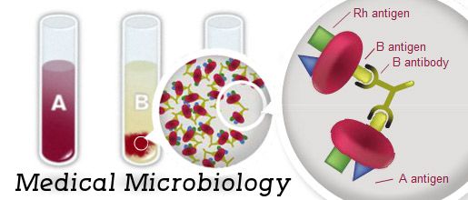
  antigen-antibody reaction is a special chemical reaction between antibodies and antigens during immune reaction. The antigens and antibodies combine by a reaction called agglutination. It is the main reaction in the body this process  protects the body  from complex foreign molecules, like pathogens and antigens. In the blood, the antigens are specifically and with more affinity combine by antibodies to make an antigen-antibody complex.  then it is  transported to cellular systems . the cellular system can  destroy or deactivate this complex. 

Application

Antigen-antibody interaction is usually used in laboratory techniques for serological test of blood compatibility and also various pathogenic infections. The most using is in  ABO blood group determination, that is helpful for blood transfusion. it also uses in  applications include ELISA,immunofluorescence, and immunoelectrophoresis.

Precipitation reaction

Soluble antigens interact with soluble antibodies in presence of an electrolyte at optimum temperature and pH to form insoluble visible complex. This is named a precipitation reaction. It is often used for qualitative and quantitative determination of antigen and antibody. It includes the reaction of soluble antigen with soluble antibodies and form large interlocking aggravated which called lattice. It happens in two stages. Firstly, the antigen and antibody rapidly form antigen-antibody complexes in few seconds then it followed by a slower reaction in which the antibody-antigen complexes forms lattices which precipitate from the solution.

A ring test is useful for diagnosis of anthrax and detection of adulteration in food.

Agglutination reaction

It happens when antigen-antibody reaction  cross-link particulate antigens and results in the visible clumping of the particle. There are two types active agglutination and passive agglutination. They are usually used in blood tests for diagnosis of enteric fever.


2020-10-17

Antibiotic sensitivity-test and result

antibiotic sensitivity test

Antibiotic sensitivity-test and result

Antibiotic sensitivity test


Antibiotic resistance

 is understood as the power of bacterium to resist antibiotics because of presence of resistance factor in its genetic material. This factor is also present usually inherited or  it may be acquired through horizontal chromosomal mutation events like conjugation, transformation, and transduction. It happens by inactivation of the porin channel, modification of antibiotic targets, and neutralizing antibiotic effectivity through enzymatic action. Therefore changes in bacterium are important to understand the resistance mechanism


Susceptibility test

 is used to see that antibiotics can inhibit the expansion of the bacterium or fungi causing a particular infection. The results from this test can facilitate a health care professional confirm that medicine are possible to be the best in treating somebody's infection

Some infections could need testing as a result of the bacterium or fungi isolated from an infection site are identified to own unpredictable susceptibility to the drugs usually used to treat them. This bacteria like staphylococci ("staph") and Pseudomonas aeruginosa

Sometimes there is also more than one kind of microorganism isolated from an infected site, like a wound infection. susceptibility testing is also used to confirm that antibiotic or antibiotic mixtures are going to be best in treating all the various varieties of bacteria causing the infection

Test purpose

To guide the practitioner in choosing the most effective antibiotic agent for a  patient

To management the utilization of inappropriate antibiotics in clinical observe

To accumulate epidemiological data on the resistance of microorganisms of public health importance at intervals the community


Type

Qualitative

For the testing of isolates from “healthy” people with intact immune defenses

For less serious infections like uncomplicated urinary tract infections

Quantitative

In the treatment of significant infections like endocarditis or osteomyelitis

For infections in risky patient groups like immunocompromised patients (e.g.. transplant patients)

Antibiotic Sensitivity Tests

Diffusion
 
Kirby-Bauer technique
 
Stokes method
 
Dilution
 
Tube Dilution
 
Agar Dilution

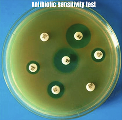
 Disk Diffusion Method

Principle

A paper disk with an outlined quantity of antibiotic is used to get a dynamically changing gradient of antibiotic concentrations within the agar within the locality of the disk

The antibiotic contained in a reservoir is allowed to diffuse out into the medium and act during a plate freshly seeded with the test organisms

The disk is put to the surface of an agar plate inoculated with the test bacteria

The antibiotic diffuses out of the disk to make the gradient

The test organism starts to divide and grow and progresses toward a crucial mass of cells


Inhibition zone edge is made at the crucial time wherever a selected concentration of the antibiotic is simply ready to inhibit the organism before it reaches an overwhelming cell mass or crucial mass

Mueller-Hinton Agar

Medium containing beef infusion, peptone, and starch

Used primarily for the disk-diffusion technique 

Mueller-Hinton agar is taken into account the most effective for routine susceptibility testing of nonfastidious bacterium 

Why

It shows acceptable batch-to-batch reliability for susceptibility testing

It is low in some antibiotics like trimethoprim, sulfonamides,  and tetracycline inhibitors

It provides satisfactory growth of most nonfastidious pathogens

A massive body of information and skill has been collected regarding status tests performed with this medium

Antibiotic Disks

Any commercially offered discs with the correct diameter and efficiency are often used

On removal from the refrigerator, the containers ought to be left at room temperature for about one hour to permit the temperature to equilibrate

Measurement of diameter

Using a ruler

 under the surface of the plate which contain transparent medium

Using a pair of calipers

on the plate which contain opaque medium

Using automatic zone readers

BIOMIC

Aura

Protozone


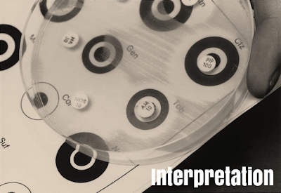
interpretation
Result interpretation

Susceptible,When the edge of the inhibition zone present outside the black circle

Resistant, When there's no zone, or once it lies at intervals the white circle

Intermediate, When the edge of the inhibition zone lies on the black circle


Using a ruler

The diameter of inhibition zone is usually  measured by using a ruler or a pair of calipers

This diameter is interpreted per the critical diameters

Factors influencing size of zone

Inoculum density

Too light substance, Inhibition zones are going to be larger even if the sensitivity of the organism is unchanged. Relatively resistant strains is also incorrectly reported as susceptible

Too significant heavy, Inhibition zones are going to be smaller. Relatively susceptible strains could then be incorrectly reported as resistant

Temperature of incubation

If the temperature is lowered , the time needed for effective growth is extended and bigger zones result

Potency of antibiotic disks

If the effectivity of the drug becomes low due to deterioration throughout storage, the inhibition zone can show a corresponding decrease in size 

Dilution ways

Used to see the lowest concentration of antibiotic to inhibit or kill the organism

occurred by dilution of antibiotic in  agar or broth media 

Minimum inhibitory concentration

The lowest concentration of drug that inhibits the expansion of the bacterium isolated from the patient

The MIC is detected by inoculating the organism isolated from the infected person into a series of tubes or cups which contain progressive dilutions of the drug


Minimum bactericidal concentration

The lowest concentration of drug that kills the microorganism isolated from the patient

E-Test

Epsilometer test

Quantitative technique of antibiotic sensitivity testing

Applies each dilution of antibiotic and diffusion of antibiotic into the medium

Combines the principles of disk diffusion and agar dilution ways

A predefined stable antibiotic gradient is present on a thin inert carrier strip

Using innovative dry chemistry technology

E-Test is used to see the on-scale Minimum inhibitory Concentration (MIC)


E-Test Uses


Determining the MIC of fastidious, slow-growing or nutritionally deficient micro-organisms, or for a particular kind of patient or infection

:Detecting

–Glycopeptide-resistant Enterococci (GRE)

–Glycopeptide-intermediate S. aureus (GISA)

–Resistant mycobacterium tuberculosis

–Extended spectrum beta lactamases (ESBL)

Detecting low levels of resistance

Testing an antibiotic not performed in routine use or a replacement, recently introduced antibiotic agent

Advantages

Simple

Accurate

Reliable